- Shopping, made easy.
- /
- Get the app!
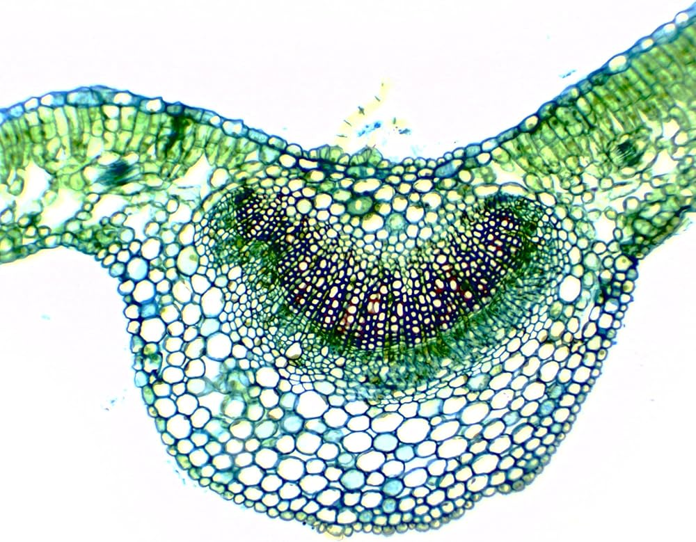
This prepared microscope slide features a cross section of a dicot leaf, providing a detailed look at plant tissue organization and vascular structure. Professionally stained to improve contrast, it clearly displays the epidermal layer, palisade and spongy mesophyll, and vascular bundles, supporting instruction in leaf anatomy and plant function.
Mounted on a 25 x 77mm (1 x 3 in.) pre-cleaned glass slide with ground edges, this specimen is a valuable tool for biology classrooms studying photosynthesis, transport, and structure-function relationships. Aligned with Next Generation Science Standards (NGSS), this slide enhances hands-on learning in plant science and comparative botany.
 10PK Cork Stoppers, Size #9-18mm Bottom, 24mm Top, 29mm Length - Tapered Shape, Natural Bark Material - Great for Household & Laboratory Use - Eisco Labs
SAR 75
10PK Cork Stoppers, Size #9-18mm Bottom, 24mm Top, 29mm Length - Tapered Shape, Natural Bark Material - Great for Household & Laboratory Use - Eisco Labs
SAR 75
 2 Prong, Vinyl Coated, Burette, Flask, Beaker Clamp on Stainless Steel Rod - 4.25" Max Clamp Opening, 5.5" Rod - Powder Coated Zinc Clamp - Research, Industrial Laboratory Grade - Eisco Labs
SAR 89
2 Prong, Vinyl Coated, Burette, Flask, Beaker Clamp on Stainless Steel Rod - 4.25" Max Clamp Opening, 5.5" Rod - Powder Coated Zinc Clamp - Research, Industrial Laboratory Grade - Eisco Labs
SAR 89
 Rubber Bulb, 1mL - Heavy Weight Rubber - for use with Pipettes & Medicine Droppers, 5-6mm in Diameter - Eisco Labs
SAR 36
Rubber Bulb, 1mL - Heavy Weight Rubber - for use with Pipettes & Medicine Droppers, 5-6mm in Diameter - Eisco Labs
SAR 36
 Serological Pipette, 1ml - Class A, Tolerance ±0.007ml - Blue Graduations - Color Code, Yellow - Calibrated for Delivery to Jet - Borosilicate Glass - Eisco Labs
SAR 60
Serological Pipette, 1ml - Class A, Tolerance ±0.007ml - Blue Graduations - Color Code, Yellow - Calibrated for Delivery to Jet - Borosilicate Glass - Eisco Labs
SAR 60