- Shopping, made easy.
- /
- Get the app!
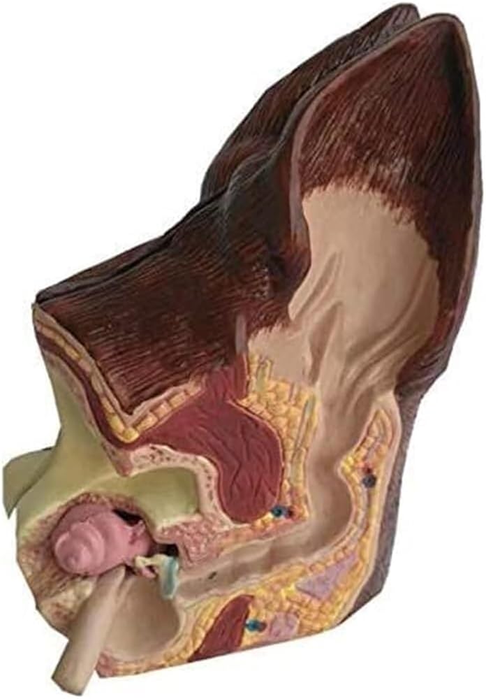
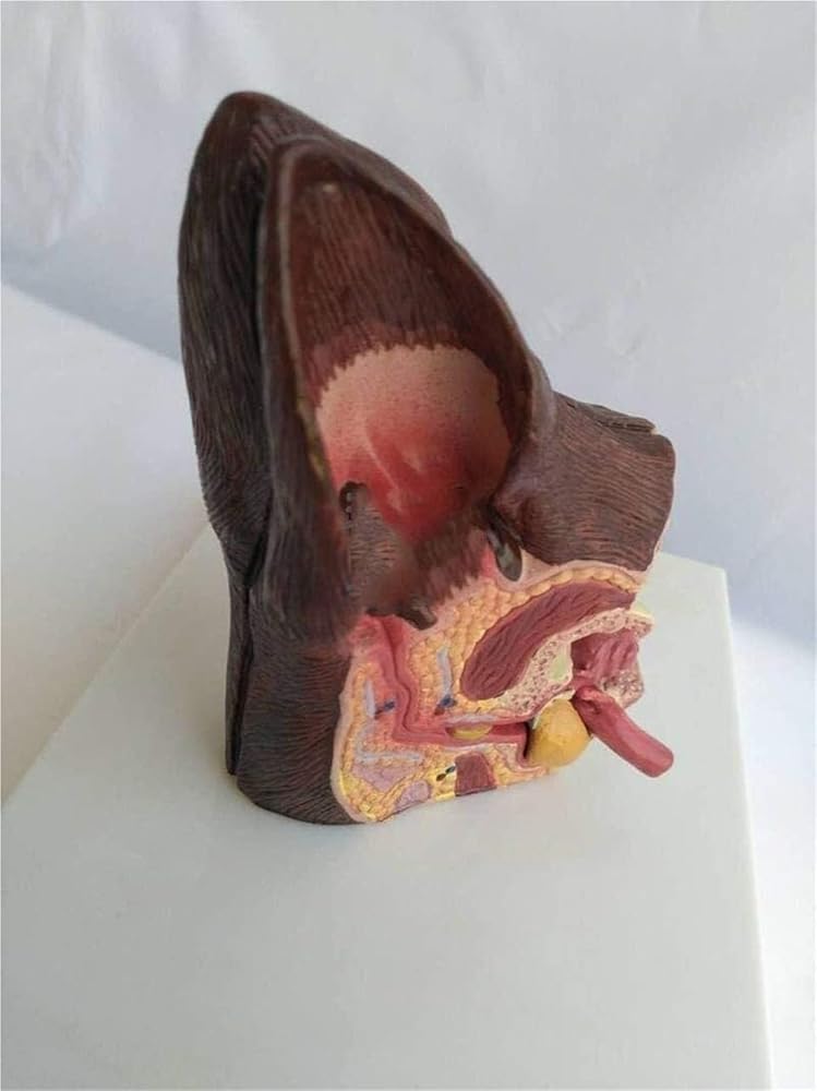
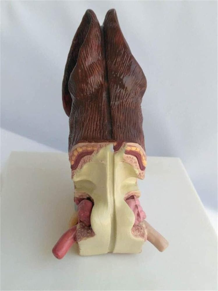
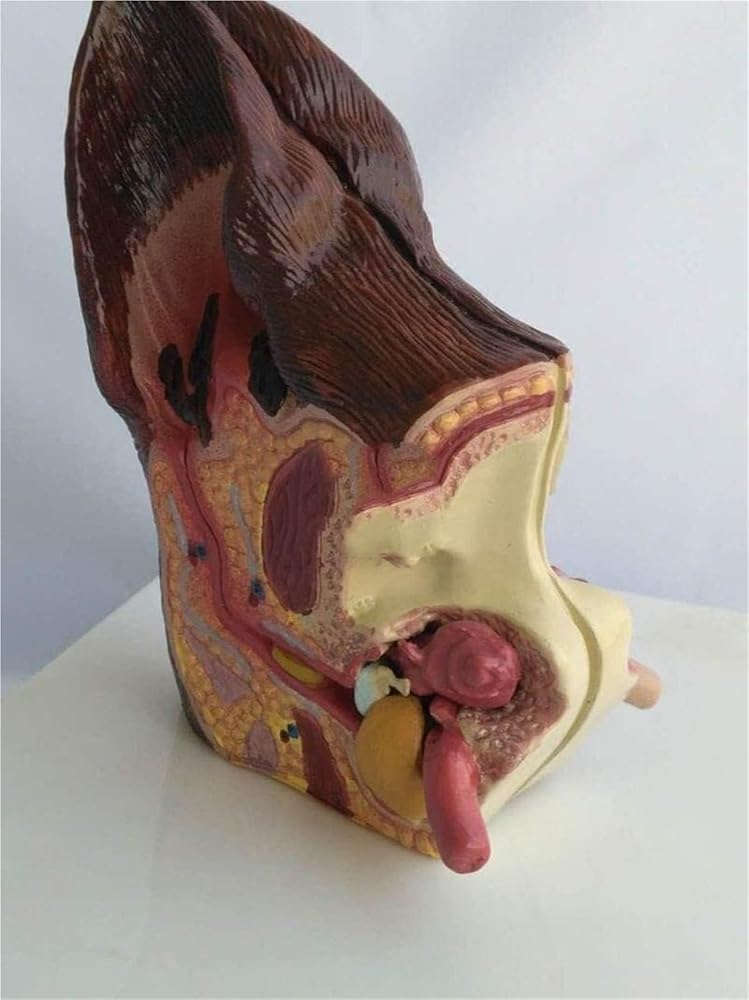
Durable plastic mold
Accurately dissect both sides: normal and diseased
Product Name: Dog Ear
Product size: 16.5 * 13 * 16CM/6.5 * 5.1 * 6.3in
Product Material: PVC
Packaging size: 1808 * 15 * 8.5CM
Weight: 0.4kg
 Multifunctional Patient Care Simulator, Human Anatomy Model for Nursing Medical Training with 24 Basic Nursing Operations
SAR 1,822
Multifunctional Patient Care Simulator, Human Anatomy Model for Nursing Medical Training with 24 Basic Nursing Operations
SAR 1,822
 Dissecting The Cricoid Membrane, Puncturing The Cricoid Cartilage, Tracheal Intubation, Suturing, and Emergency Surgical Training
SAR 2,179
Dissecting The Cricoid Membrane, Puncturing The Cricoid Cartilage, Tracheal Intubation, Suturing, and Emergency Surgical Training
SAR 2,179
 Anatomical Model of Human Shoulder Joint Muscles
SAR 1,472
Anatomical Model of Human Shoulder Joint Muscles
SAR 1,472
 Full Semester Delivery of Models for Human Anatomy Learning, Presentation, and Teaching in Medicine
SAR 3,786
Full Semester Delivery of Models for Human Anatomy Learning, Presentation, and Teaching in Medicine
SAR 3,786