- Shopping, made easy.
- /
- Get the app!
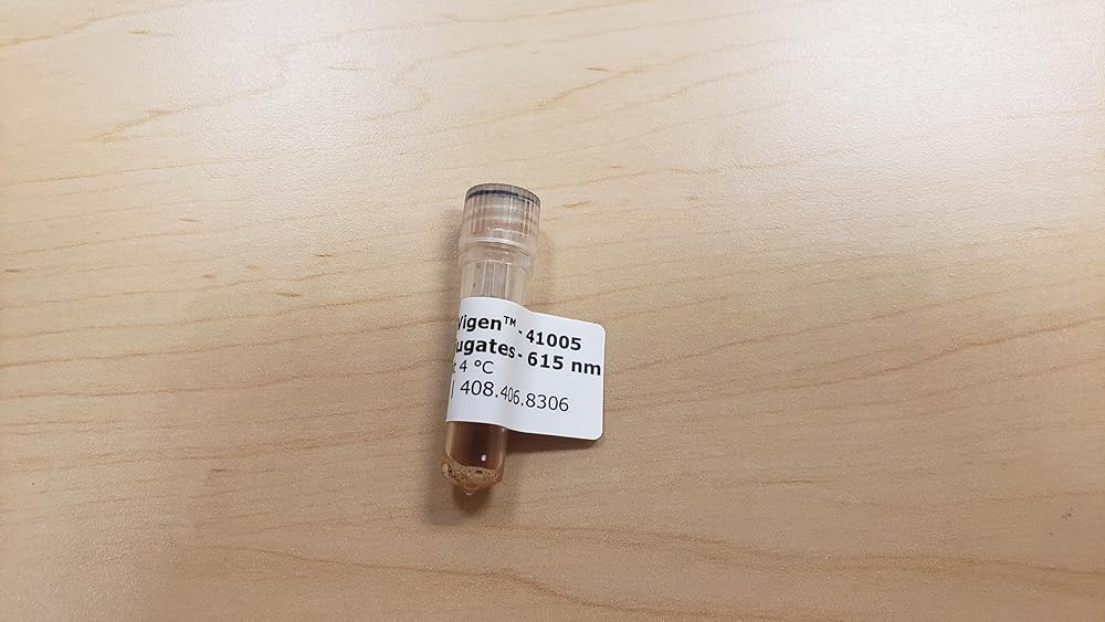
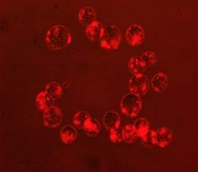
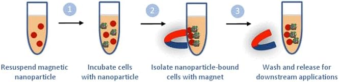
MyQuVigen nanoparticles represent a breakthrough in cell separation technology, combining superparamagnetic iron oxide with quantum dots for superior performance. These fluorescent magnetic nanoparticles feature streptavidin conjugates with peak emission at 615 nm, offering bright, stable fluorescence and strong magnetic properties. The particles can be excited at 488 nm or shorter wavelengths, making them versatile for various imaging applications. Perfect for cell isolation and labeling, these nanoparticles enable column-free magnetic separation, preserving cell viability for downstream analysis. The streptavidin coating allows universal binding to biotin-conjugated antibodies with high specificity and binding capacity. Researchers can achieve consistent, high-quality results in cell separation and fluorescence imaging applications. The nanoparticles are ideal for isolating specific cell populations like CTCs and stem cells, with the separated cells remaining viable for further culture or molecular analysis. The strong fluorescent signal enables direct microscope imaging and fluorescence-based cell analysis without additional processing steps.
 Edvotek 101 Electrophoresis Agar 6 Kit, 6-Gel, 1" Height, 1" Wide, 1" Length
SAR 219
Edvotek 101 Electrophoresis Agar 6 Kit, 6-Gel, 1" Height, 1" Wide, 1" Length
SAR 219
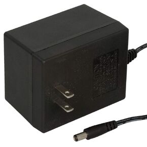 Jameco ReliaPro 24 Volt AC 2000 mA 48 Watt Linear Wall Adapter 2.5mm Plug
SAR 107
Jameco ReliaPro 24 Volt AC 2000 mA 48 Watt Linear Wall Adapter 2.5mm Plug
SAR 107
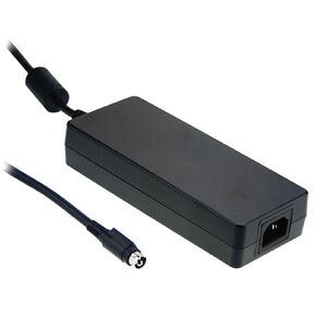 MEAN WELL GST160A24-R7B 160W 24V 6.67 Amp AC-DC High Reliability Industrial
SAR 278
MEAN WELL GST160A24-R7B 160W 24V 6.67 Amp AC-DC High Reliability Industrial
SAR 278
 Jameco Valuepro DDU120050F0981 AC-to-DC Regulated Linear Wall Adapter, 12V, 0.5 Amp, 6W
SAR 78
Jameco Valuepro DDU120050F0981 AC-to-DC Regulated Linear Wall Adapter, 12V, 0.5 Amp, 6W
SAR 78